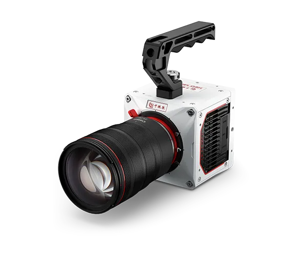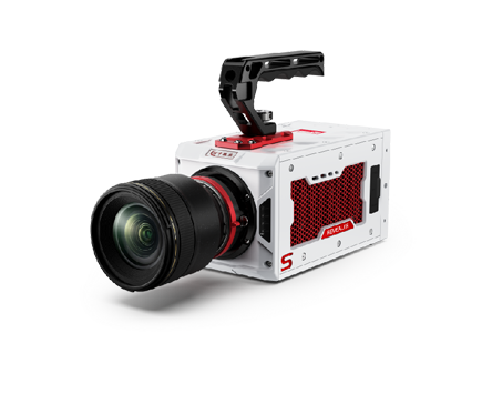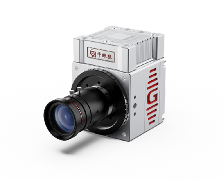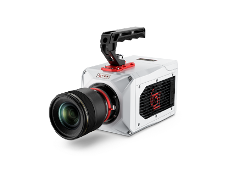
A Revealer Gloria 4.2 sCMOS scientific camera combined with a fluorescence microscope was used to image the spatial distribution of weak fluorescence signals in an oil-and-gas microfluidic chip. The experiment focused on detecting fluorescently labeled oil within micro/nanochannels under low-light conditions, providing image data for subsequent analysis of fluid distribution in microfluidic structures.
Microfluidic chips provide a visualized microscale platform for studying fluid transport in confined geometries and complex pore structures. In this experiment, the objective was to use a high-sensitivity scientific camera to detect weak fluorescence signals in micro/nanochannels and identify the spatial distribution of fluorescently labeled oil within the channels.
The main imaging challenges were the low fluorescence intensity inside the micro/nanochannels, nonuniform illumination from the LED fluorescence source, and stray light that further reduced the contrast between the target signal and background. To address these issues, the research team at China University of Petroleum introduced a Revealer sCMOS camera together with a low-light imaging strategy to improve the visibility of weak fluorescence signals for spatial distribution analysis.
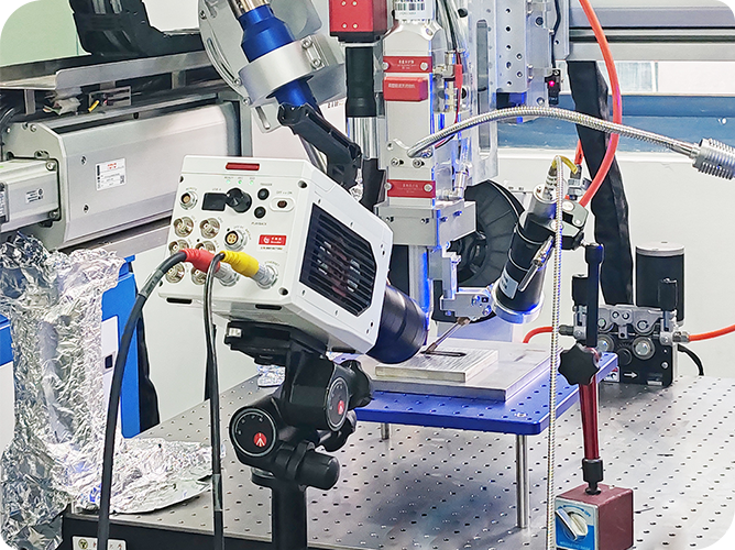
The microfluidic scientific imaging system consisted of a Revealer Gloria 4.2 scientific camera, a fluorescence microscope, and an LED fluorescence light source. The smallest channel structures in the microfluidic chip had a depth of approximately 100 nm and a width of approximately 1 μm.
The main sCMOS camera settings used in the experiment included:
2×2 binning;
maximum exposure time of 4 s;
CMS / low-noise imaging mode;
dark-field correction;
flat-field correction.
The Revealer sCMOS scientific camera converted weak fluorescence signals reaching the detector into digital images suitable for subsequent analysis.
A fluorescence microscope was used to observe the microfluidic chip under 6× and 20× magnification conditions, with an LED fluorescence source used for illumination.
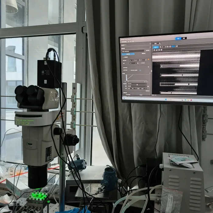
Figure 1. Revealer Gloria 4.2 sCMOS camera and fluorescence microscope system for weak fluorescence imaging in microfluidic micro/nanochannels.
The experimental sample was an oil-and-gas microfluidic chip with minimum channel dimensions of approximately 100 nm in depth and 1 μm in width. Both fluorescence and bright-field signals were observed, with particular attention paid to the spatial distribution formed after fluorescently labeled oil entered the micro/nanochannels. Fluorescence labeling converted the spatial occupancy of the fluid inside the transparent microfluidic chip into measurable grayscale differences in microscopic images.
Exposure conditions were adjusted according to the imaging task. For dynamic observation of oil flow, an exposure time of approximately 350 ms was used, whereas the exposure time was extended to 4 s for static fluorescence distribution imaging to accumulate more weak fluorescence signal. The experiment also used 2×2/4×4 binning and the CMS low-noise imaging mode to improve image visibility under low-signal conditions. Dark-field and flat-field correction were applied to reduce the influence of fixed background signals and nonuniform LED illumination. The resulting scientific images were used to identify the spatial fluorescence distribution within the micro/nanochannels, followed by fluorescence-region extraction and labeling using the ImageJ Threshold function.
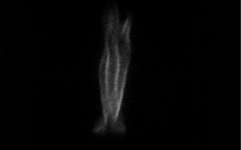
After optimization of the low-light imaging parameters, the micro/nanochannels were clearly illuminated in the acquired images, indicating that fluorescently labeled oil had entered the corresponding channels. The smallest channel structures in the chip had a depth of approximately 100 nm and a width of approximately 1 μm.
In this microfluidic experiment, the primary value of the sCMOS camera was to convert previously difficult-to-observe weak fluorescence signals into scientific images suitable for analysis, allowing researchers to identify the spatial distribution of oil within the micro/nanochannels.
It should be noted that the 100 nm value refers to the structural depth of the microfluidic channel rather than the optical spatial resolution of the imaging system.
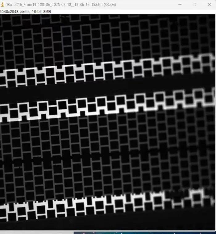
Figure 2. Weak fluorescence image of microfluidic micro/nanochannels captured with a Revealer Gloria 4.2 sCMOS camera at 4-second exposure.
The fluorescence images were further processed using the ImageJ Threshold function to identify and mark regions occupied by fluorescently labeled oil. The original grayscale microscopic images could therefore be converted into fluorescence distribution data with defined spatial boundaries, providing an imaging basis for subsequent analysis of channel occupancy, spatial distribution differences, and time-resolved transport processes.
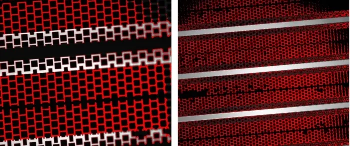
Figure 3. ImageJ Threshold analysis of microfluidic fluorescence images acquired with a Revealer Gloria 4.2 sCMOS scientific camera.
1) A Revealer Gloria 4.2 sCMOS scientific camera combined with fluorescence microscopy was used for weak fluorescence imaging of a microfluidic chip. Long exposure, low-noise imaging, 2×2/4×4 binning, dark-field correction, and flat-field correction improved the visibility of weak fluorescence signals under nonuniform LED illumination and stray-light conditions.
2) The results showed that the spatial distribution of fluorescently labeled oil within the micro/nanochannels could be clearly recorded and further extracted using ImageJ Threshold processing.
3) For microfluidics research, the value of an sCMOS camera is not simply to increase image brightness, but to convert weak optical signals under low-photon-flux conditions into observable and analyzable scientific imaging data. This provides an experimental basis for studying fluid distribution and subsequent transport processes in micro/nanochannels.
 English
 Deutsch